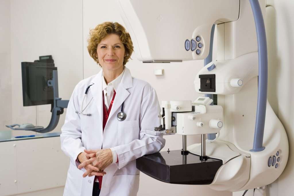
Roughly 1 in 8 women in the United States develop breast cancer in their lifetime. * As a patient, friend or family member, nearly everyone is affected by this disease in some way.
Rebecca Lewis, MD, primary care physician with St. Mary’s Physician Associates, talks about the importance of screenings for early detection of breast cancer.

Why is early detection of breast cancer so important?
Regular breast cancer screenings are important because they can detect breast cancer years before symptoms develop. Early detection is key, because success rates are much higher when breast cancer is detected and treated during its early stages. A visit with your primary care physician is a good first step in breast health care because he or she can provide guidance on what screenings may be appropriate for your personal health.
What types of screenings are important?
There is a comprehensive range of imaging and biopsy services available including:
• 2D and 3D digital mammography (breast cancer detection)
• Invenia™ Automated Breast Ultrasound System (ABUS) system (breast cancer detection)
• Bone density assessment (for fracture risk due to osteoporosis)
• Breast biopsy (ultrasound, fine needle, stereotactic)
• Resources for breast cancer patients

How effective is digital mammography?
Mammograms are the most widely used imaging method for detecting breast cancer, often spotting issues before anything can be felt. Low-radiation digital mammograms are very effective, identifying upwards of 85% of all abnormalities in the breast. The American Cancer Society strongly advises women who are age 40 and older to have a yearly mammogram. Individuals who are at higher risk of breast cancer, such as those with a family history of breast cancer or history of high-dose radiation exposure prior to the age of 30, may require screening earlier than 40. If you believe you are at a higher risk, make sure to talk to your primary care physician about when to begin screenings.
St. Mary’s Women’s Imaging Center also offers 3D mammography, which is also called tomosynthesis digital mammography. 3D mammography is different from standard mammography because it takes multiple images of the breast at various levels and provides more detail. This can help make it easier to identify abnormalities and is particularly useful for evaluating dense breast tissue. It can also reduce the number of callbacks for repeat testing.
What does having dense breasts mean and what screening is needed for this?
Dense breasts have a higher proportion of glandular and connective tissues as compared to fatty tissue. Women who have been told they have dense breasts should be aware that it can make it difficult for screening mammography to detect tumors. What’s more, women who have dense breasts are also at increased risk for breast cancer.
Additional imaging tests – ultrasound, MRI or molecular breast imaging – may be needed for complete evaluation.
What is breast ultrasound?
Ultrasound is a noninvasive, non-radiation examination that uses sound waves to detect disease and locate possible abnormalities in breast tissue. It is designed to provide doctors with precise images for efficient diagnosis of breast problems, and can be effective in distinguishing certain abnormalities in the breast such as lumps, solid masses and cysts. The systems enable the physician to perform high-resolution panoramic imaging or 3D scanning in real time.
The Women’s Imaging Center is the first provider in the Northwestern Oklahoma and Southern Kansas regions to use the Invenia Automated Breast Ultrasound System (ABUS) for breast cancer screening. This system is used in addition to mammography for asymptomatic women with dense breast tissue and no prior interventions. ABUS can help improve detection of small cancers in dense breast tissue that cannot be seen on a mammogram alone.

When would a breast biopsy be necessary?
Breast biopsies are used to check suspicious or unusual areas in breast tissue for cancerous cells. The newest procedure, vacuum-assisted breast biopsy, uses a minimally invasive system in which the doctor uses mammography (stereotactic-guided biopsy) or ultrasound to locate the suspicious area. He or she then makes a tiny incision in the breast and uses a small probe with a vacuum to gently draw, cut and collect tissue into the probe’s hollow chamber.
To make an appointment with Dr. Lewis, call 580-233-5553. To view the provider directory, visit stmarysphysicianassociates.com. To learn more about our imaging services, call 580-249-3930 or visit stmarysregional.com/imaging.
Physicians are on the medical staff of St. Mary’s Regional Medical Center, but, with limited exceptions, are independent practitioners who are not employees or agents of St. Mary’s Regional Medical Center. The hospital shall not be liable for actions or treatments provided by physicians. For language assistance, disability accommodations and the nondiscrimination notice.
*American Cancer Society (ACS)


